Many cellular mechanisms have been linked to autism, including changes in mitochondria and synapses.
Focusing on mitochondria, Zhenglong Gu of Cornell University and the Greater Bay Area Institute of Precision Medicine, Fudan University, China, presented his work examining genetic mutations in mitochondrial DNA and a new methodology he developed to sequence mitochondrial DNA at a single cell level. Mitochondria contain their own DNA but do not house the repair mechanisms that normally safeguard nuclear DNA against mutation. Gu proposed that an accumulation of mutations to mitochondrial DNA during life could contribute to a cell’s decline and alter function of the immune system, epigenetic regulation and stem cell regeneration. Findings from his work, which he has so far conducted in Huntington’s disease, show that pathogenic mitochondrial DNA mutations come to predominate with time and signal a progressive decline of mitochondrial function. He is also working on mitochondrial DNA mutations in ASD.
Hisashi Umemori of Boston Children’s Hospital presented his efforts to understand how PCDH19, which encodes a brain-specific cell adhesion molecule, works in regulating synaptic development. Typically the PCDH19 protein is expressed at points of contact between neurons; when extracellular domains of PCDH19 on different neurons bind to each other, this stabilizes the contacts. PCDH19 molecules also bind to and cluster another cell adhesion molecule, N-cadherin, which kicks off intracellular changes important for synapse formation.
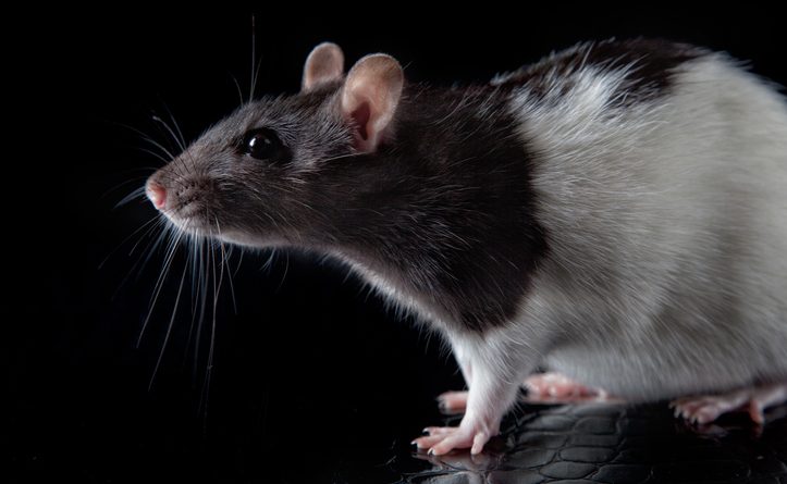
PCDH19 mutations in humans result in a disorder with seizures, cognitive difficulties and autism; though PCDH19 is located on the X chromosome, the disorder is only found in heterozygous females, not hemizygous males. Umemori presented recent experiments in mouse models that suggest an explanation for this puzzle, which involves a mismatch between PCDH19 expression across cells in the female heterozygous mice. When PCDH19 is missing on one side of a contact between neurons in the hippocampus, N-cadherin is unable to cluster appropriately, resulting in a disruption of N-cadherin signaling and mossy fiber synaptic development; when PCDH19 is missing from all cells, as it happens in the hemizygous male mice, however, N-cadherin can now bridge across contacts by themselves and cluster to kick start synapse formation. Over-expressing N-cadherin could also rescue synaptic function in female mice with a Pcdh19 mutation, which suggests a therapeutic strategy.
Immature spine development in the brain also marks fragile X syndrome. Hye Young Lee of the University of Texas Health Science Center at San Antonio has been focusing on the role of microglia, the resident immune cells in the brain, in pruning away spines. Lee has found that microglia-specific removals of FMR1, the gene mutated in fragile X syndrome, recapitulated the increased spine density and immature spine shapes found in fragile X mouse models in both cortex and hippocampus. She is now examining these mice for autism-associated behavioral phenotypes, such as repetitive behaviors and learning and memory tasks. Experiments with microglia in culture find that those without FMR1 are more reactive in their immune responses, a property that may well inform the development of therapies attempting to restore FMR1 expression in microglia.