When a mouse became aroused, some neurons became more active and others less active. As the mouse transitioned back to a more restful state, the activity patterns of these two populations largely reversed. The researchers named the populations “Arousal+” and “Arousal−” neurons.
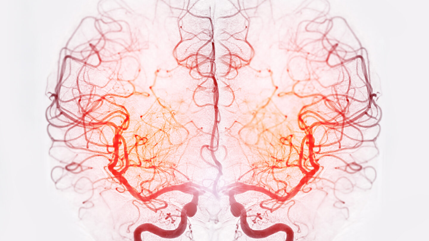
When the researchers tried to model blood flow dynamics using the bulk firing rate of all neurons together, the resulting prediction was poor, as expected. When they instead created a model with separate blood flow relationships for the two populations, their prediction improved markedly. It remained accurate as the team examined different brain regions and mental states. The variations that previous studies had found across brain regions are explained, the researchers propose, by the fact that different regions have different proportions of Arousal+ and Arousal− neurons.
“This shows that the relationship between neural activity and blood volume is more complicated than people thought, but it’s fixed across brain regions and states,” Carandini says. “We can go from neural activity to blood volume in a rock-solid way.”
A New Challenge for fMRI
What’s harder is to go in the other direction, from blood volume to neural activity. Neural activity, the new study shows, is inherently two-dimensional, incorporating separate information from Arousal+ and Arousal− neurons. It’s clear how to collapse this two-dimensional signal into a one-dimensional blood volume signal. But there’s no simple way to go in the other direction unless you have additional information, such as the relative proportion of Arousal+ and Arousal− neurons in the brain region you are studying.
Carandini likens the situation to hearing noise from a distant sports arena and wondering what is happening in the game. If you hear a roar from the crowd, maybe the home fans are cheering and the away fans are groaning — or maybe the reverse is true. “You know that something happened, but you can’t say what,” Carandini says.
That’s bad news for neuroscientists who rely on fMRI to gain insight into neural activity. “The fMRI researchers would like to just look at blood volume and infer what neurons are doing,” Carandini says. “We’re saying you can’t do that.”
The new work has given rise to a host of questions about the two neuronal populations. Apart from their involvement in arousal, “we don’t know what they do and what they are,” Landemard says. She plans to examine how neuromodulators such as serotonin influence the activity of Arousal+ and Arousal− neurons, in the hope of resolving some of these mysteries.
Perhaps the biggest mystery is why these two populations appear in every part of the brain, including regions such as the visual and auditory cortices, which are not traditionally associated with arousal. “It’s really bizarre that all regions have neurons that go up with arousal or down with arousal,” Carandini says. “We don’t know why, but we’re looking forward to learning more.”