Distinct Patterns of Connectivity Underlie Differences in Sensory Responsiveness in Autism
Featuring
-

Susan Bookheimer
SFARI Investigator
Bringing people and ideas together to fund, conduct, and share transformative science.
Our grants advance knowledge and strengthen connections between science and society.
Discover the latest news, research, and updates.
We convene scientists, mathematicians, and thinkers to share ideas and explore new questions.
Founded by Jim and Marilyn Simons, we’re a private philanthropy advancing mathematics and basic science.
Work with us on the frontier of basic science research.
The Flatiron Institute is a research center that uses computational science to advance fields like physics, biology, and astronomy.
Search
Susan Bookheimer and colleagues show that the degree of sensory overresponsivity in individuals with autism may be driven by distinct patterns of connectivity between the prefrontal cortex and amygdala.
SFARI Investigator
Sensory overresponsivity (SOR), an atypical negative reaction to sensory stimuli, is a relatively common feature of autism, although the degree of SOR can be highly variable. Previous work has shown that SOR may reflect both a reduced degree of habituation to sensory stimuli as well as reduced regulation of amygdala responses to such stimuli. Atypical but distinct neural responses may be seen in individuals with autism across the range of sensory responsiveness.
Now, SFARI Investigator Susan Bookheimer and colleagues have used functional magnetic resonance imaging (fMRI) to deepen our understanding of atypical neural responses underpinning SOR in autism, with possible implications for the design of interventions.
In the new work — partly funded by a SFARI Pilot Award — the team carried out an fMRI study of 42 children and adolescents with autism, as well as 27 typically developing control subjects. Individuals with autism were divided into high-SOR and low-SOR groups. Brain responses were measured in somatosensory, auditory, visual and prefrontal cortices, as well as amygdala, in response to joint auditory and tactile stimuli across six blocks of time.
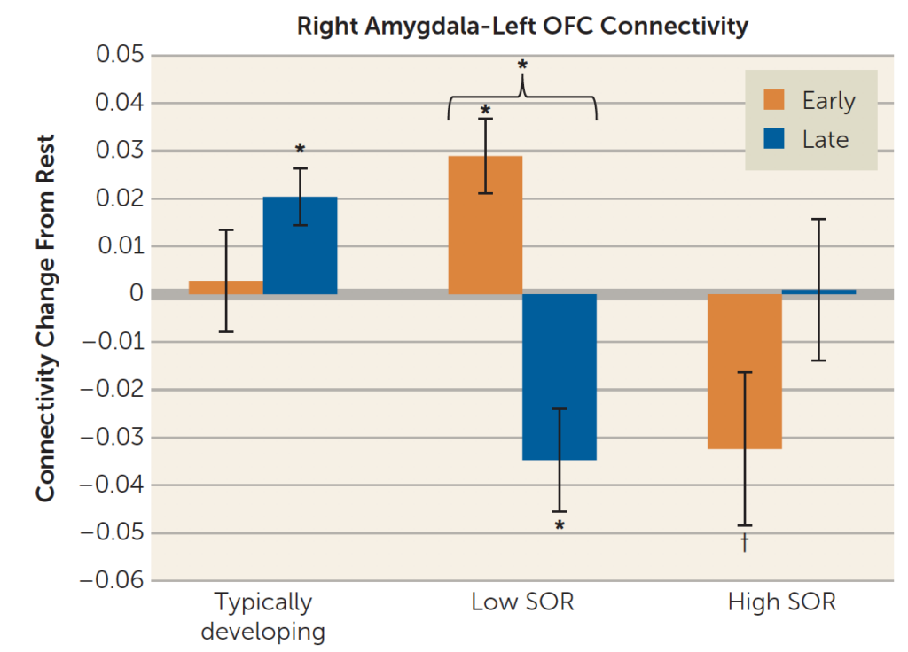
They observed that the key SOR group difference is in the pattern of connectivity across sensory exposure: the high-SOR group showed initial but unsustained amygdala-prefrontal connectivity, whereas the low-SOR group switched from positive to negative amygdala-prefrontal connectivity across sensory exposure. Moreover, the low-SOR group uniquely lacked neural responsiveness to new but similar stimuli, suggesting a degree of overgeneralization.