Dissecting the inner workings of a cell requires imaging methods with molecular specificity, molecular-scale resolution and dynamic imaging capability such that molecular interactions inside the cell can be directly visualized. Fluorescence microscopy is a powerful imaging modality for investigating cells largely owning to its molecular specificity and dynamic imaging capability. However, the spatial resolution of light microscopy, classically limited by the diffraction of light to a few hundred nanometers, is substantially larger than typical molecular-length scales in cells. Hence many subcellular structures and dynamics cannot be resolved by conventional fluorescence microscopy. We developed a super-resolution fluorescence microscopy method, stochastic optical reconstruction microscopy (STORM), which breaks the diffraction limit. STORM uses single-molecule imaging and photo-switchable fluorescent probes to temporally separate the spatially overlapping images of individual molecules. This approach has allowed multicolor and three-dimensional imaging of living cells with nanometer-scale resolution and enabled discoveries of novel sub-cellular structures. In this talk, I will discuss the technological development and biological applications of STORM.
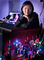
The Zhuang research lab works on the forefront of single-molecule biology and bioimaging, developing and applying advanced optical imaging techniques to study the behavior of individual biological molecules and molecular assemblies in vitro and in live cells. Students and postdoctoral fellows in the Zhuang lab apply their diverse backgrounds in chemistry, physics, biology and engineering to develop new imaging probes and methods and use these tools to study a variety of interesting biological systems. Our current research is focused on three major directions: (1) Developing super-resolution optical microscopy that allows cell and tissue imaging with nanoscopic-scale resolution and applying this technology to cell biology and neurobiology, (2) investigating how biomolecules function, especially how proteins and nucleic acids interact, using single-molecule fluorescence imaging and spectroscopy, (3) investigating how viruses and cells interact using imaging techniques with high spatiotemporal resolution.