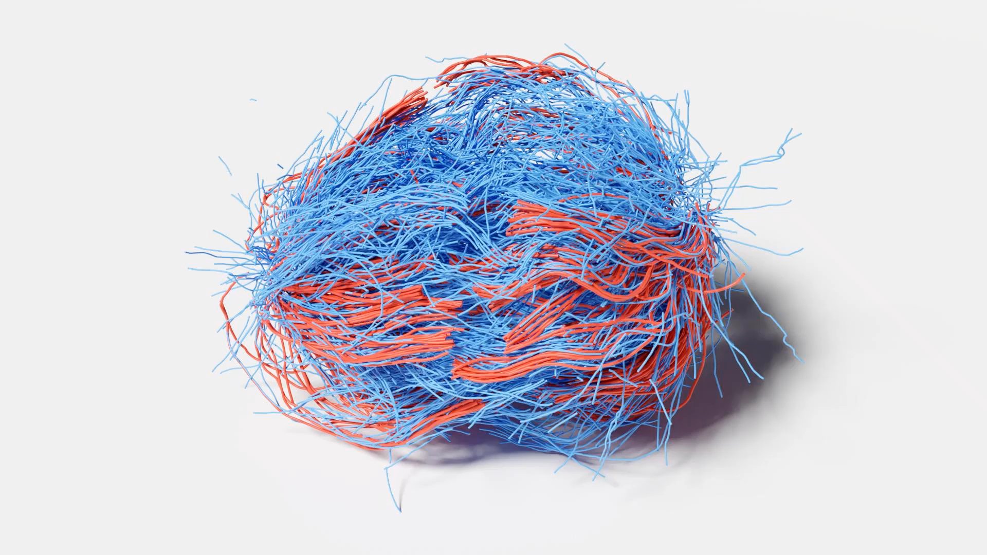
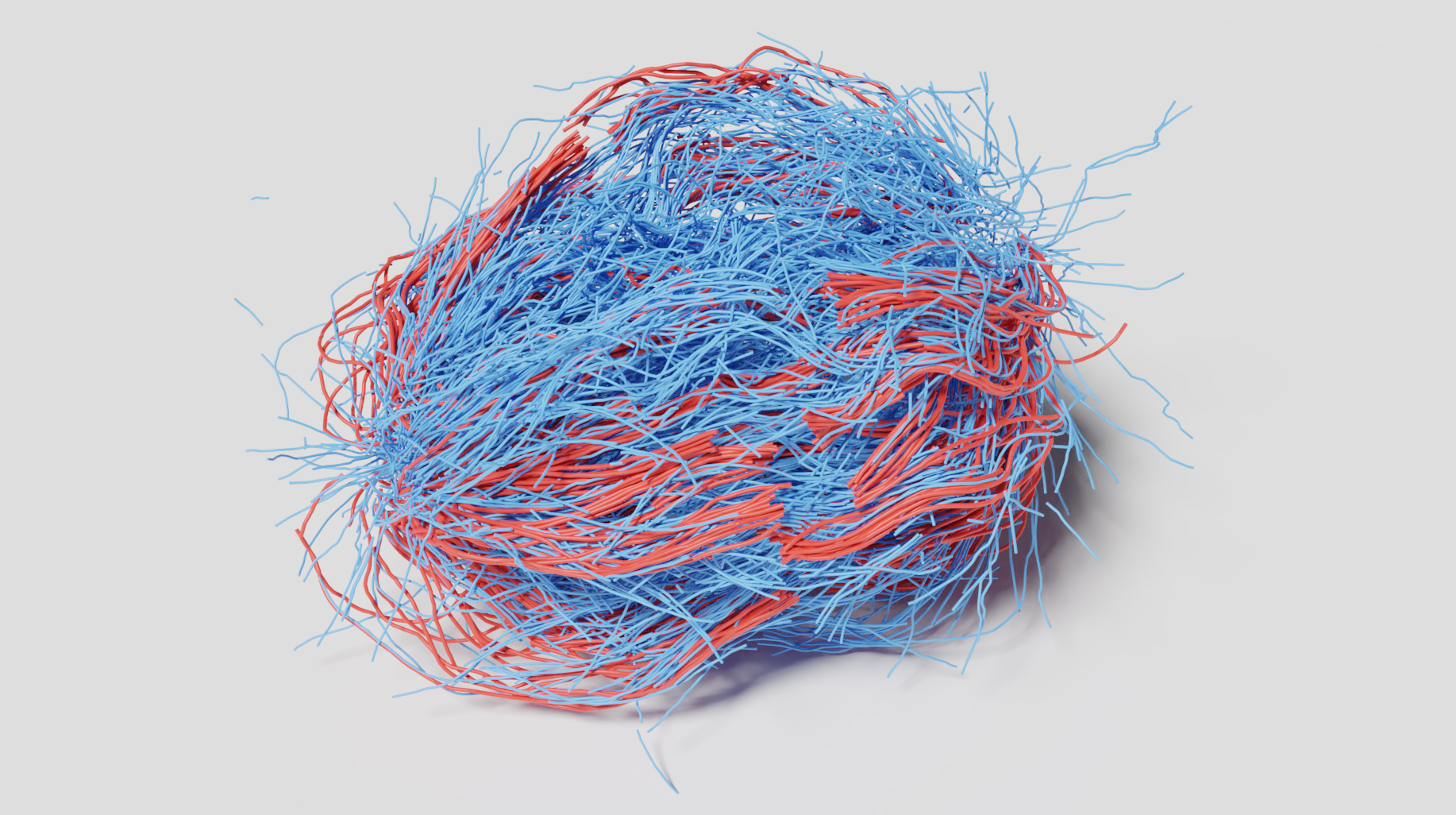
Spindles are responsible for pulling apart your chromosomes when your cells divide. New research confirms a theory that these spindles self-organize and interact with one another much like active liquid crystals. While the model successfully described the behaviors of many microtubules (blue), further research is required to capture those of kinetochore microtubules (red), which attach to chromosomes. Reza Farhadifar/Flatiron Institute
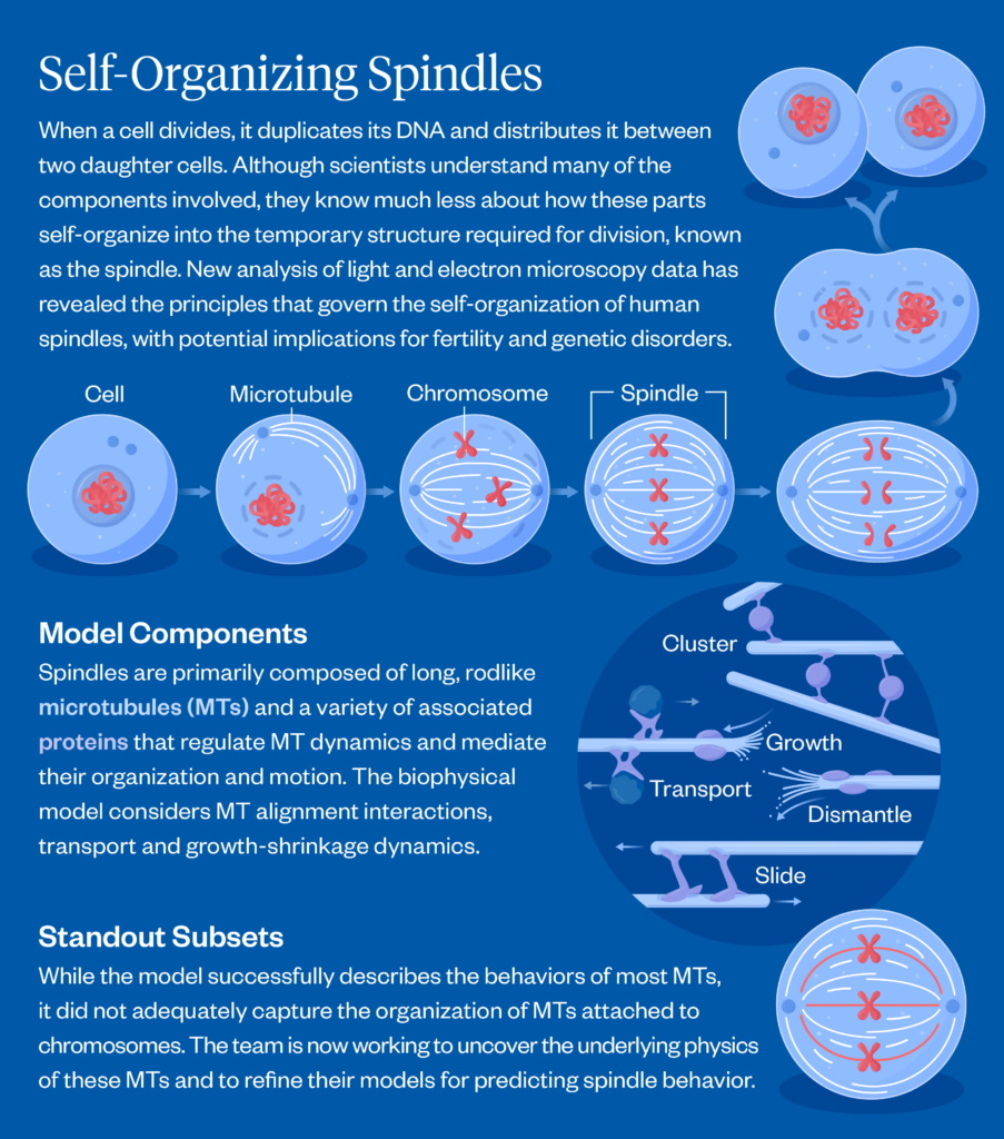
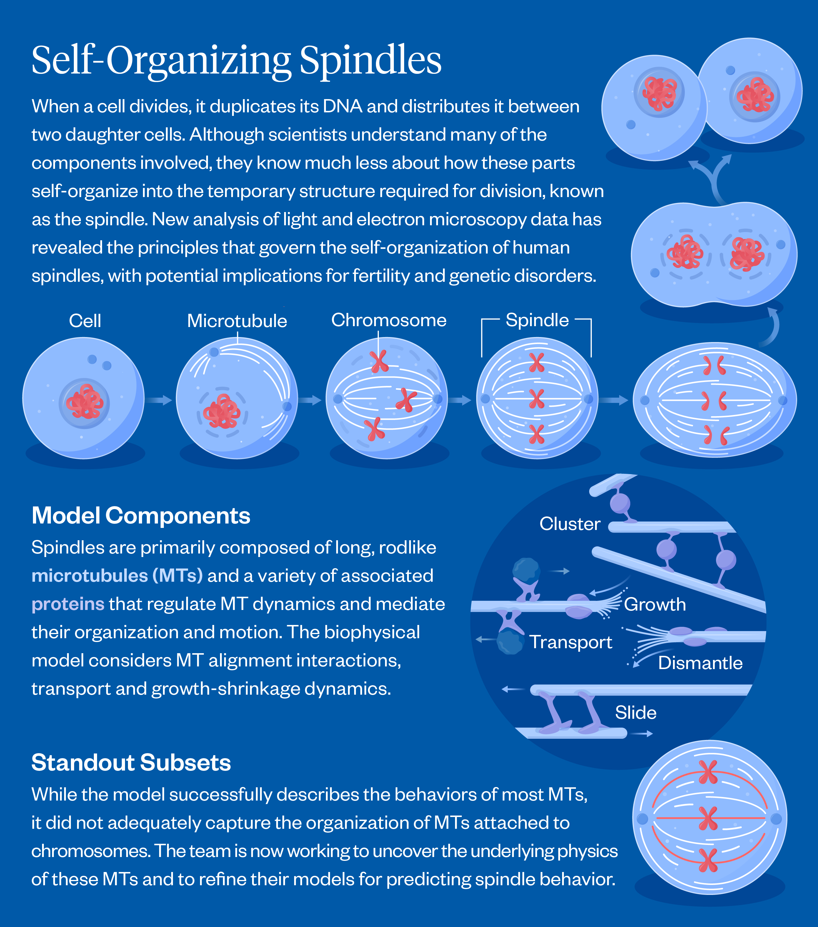
When a cell divides, it performs a feat of microscopic choreography — duplicating its DNA and depositing it into two new cells. The spindle is the machinery behind that process: It latches onto chromosomes (where DNA is stored) and separates them so they can settle into their new homes. This tricky process can sometimes go wrong, causing infertility, genetic disorders or cancer.
Scientists have a good understanding of what spindles are made of: long, thin rods called microtubules as well as a variety of associated motor proteins. However, how these microtubules interact and organize to guide the spindles’ function has remained a mystery.
One approach to understanding how the spindle self-organizes is to treat it like an active liquid crystal. Liquid crystals, like spindles, are made up of elongated subunits. Unlike liquid crystals in LCD displays, which require an external electric field to reorient their subunits, spindles are active materials that generate forces internally. Treating the spindle as an active liquid crystal enables scientists to better model interactions between microtubules, but until now, this theory had not been tested using data from actual human cells.
Drawing on data collected from dividing cells using advanced microscopy techniques, researchers at the Simons Foundation’s Flatiron Institute and their collaborators have now shown that in most cases, active liquid crystal theories hold up. They report their findings the week of February 9 in Proceedings of the National Academy of Sciences.
“The fact that we get consistent answers from two very different data sets is very encouraging,” says Suryanarayana Maddu, a first author of the paper and a Flatiron Research Fellow at the institute’s Center for Computational Biology (CCB). “It suggests the observed patterns reflect real physical properties of the spindle rather than experimental artifacts and demonstrates how theories based in physics and materials science can be applied to study biological systems.”
Their work, while rooted in basic science, could eventually inform research on fertility and in vitro fertilization (IVF), since accurate chromosome segregation is critical for egg viability. Issues surrounding the process can lead to genetic conditions such as Down syndrome.
“It’s possible that by applying quantitative biophysics approach like ours, you could analyze the spindle in a single egg cell or early embryo and measure properties that aren’t currently assessed during treatments like IVF,” says Colm Kelleher of Syracuse University, the study’s other first author. “Those properties might tell you something useful about the developmental potential of that egg or embryo.”
A better understanding of spindles could also aid cancer treatments, as many chemotherapy drugs target spindles.
“Cancer cells divide constantly, so a common strategy is to use drugs that preferentially harm dividing cells,” says Kelleher. “That’s one reason spindles are such an important focus — understanding how they work, how they fail and how we might manipulate them with drugs.”
The new study is co-authored by Harvard University’s Mustafa Basaran and Daniel Needleman (also a senior research scientist at CCB), Dresden University of Technology’s Thomas Müller-Reichert and the Flatiron Institute’s Michael Shelley. The work is part of an initiative within CCB called CCBx, which aims to unite theorists and experimentalists to develop and test theories of biological systems.
Theory Meets Experiments
Maddu and Kelleher came from applied mathematics and physics backgrounds before embarking on research in biology, and they viewed the question of spindle organization through that lens.
“One way to think about our work in this paper is that we’re extending tools from materials physics, originally developed for much simpler, inert materials like water or metals to understand this living material that spindles are made of,” says Maddu.
Testing the active liquid crystal theory experimentally is challenging. Most data on spindles come from light microscopy, which allows scientists to image living cells and track spindle dynamics over time but lacks the resolution to directly observe individual microtubules.
Another type of microscopy — called electron microscopy — can fill this gap. With electron microscopy, scientists can zero in on much tinier structures, such as individual microtubules, but they lose the ability to track the spindle dynamics over time in living cells.