
Working-Memory Research Uncovers Essential Role for Thalamus
A trio of papers published in May suggest that short-term memory depends on interactions between the cortex and thalamus, a subcortical brain region that connects to many parts of the brain. Previous research had suggested that the cortex generates and maintains the neural activity thought to underlie working memory. But the new findings show that the thalamus is essential, changing the notion of how working memory works. The results add to a growing body of evidence that the thalamus is much more complex than its simple textbook description as a relay station that ferries information from the sensory system to higher brain areas.
“It’s a pretty remarkable series of papers,” says Xiao-Jing Wang, a neuroscientist at New York University and investigator with the Simons Collaboration on the Global Brain (SCGB), who was not involved in the studies. Most previous research on working memory has focused on the cortex, specifically the prefrontal cortex, Wang says. “But all three groups found that the thalamus, a subcortical system, plays an important role in working memory — it’s not just a receiver of the signal, it’s an essential partner with the cortex.”
The new papers are also notable in that they study working memory in rodents rather than primates. Mice offer the advantage of better tools for precisely manipulating and tracking neurons, allowing scientists to more directly investigate the role that different brain regions play. But it’s only in the last few years that researchers have developed appropriate behavioral tasks for probing working memory in mice.
Working memory refers to the ability to recall a small number of items for tens of seconds. Decades of research have linked working memory to sustained activity in a subset of cortical neurons — these neurons start firing when you need to remember something, such as a phone number, and they stop firing when you punch the number into your phone.
One of the unsolved mysteries of working memory is how neurons maintain this activity for seconds. Individual neurons operate at much shorter timescales, firing on the order of milliseconds. “How do they create a persistent state that correlates with working memory?” asks Karel Svoboda, a neuroscientist at the Howard Hughes Medical Institute Janelia Research Campus and SCGB investigator. “That’s the core mystery.”
To study this question, Svoboda and collaborators trained mice in a tactile discrimination task. The animals had to move in a certain direction based on a tactile cue — the location of an object detected via their whiskers — but only after a one to two second delay. “They have to remember their decision about location and plan the movement,” Svoboda says.
Previous research suggests that the neural activity responsible for this type of memory is generated and maintained in the cortex, in this case, the anterior lateral motor (ALM) cortex. “There are lots of excitatory neurons in that area — one neuron can excite another and another, which could excite the first one, generating a ping-pong mechanism,” says Svoboda.
But Svoboda and collaborators’ new results challenge that idea. They found that cortical activity during the delay is critically dependent on activity in the thalamus, and vice versa — inhibition of firing in one region stops firing in the other. “To our surprise, the thalamus is an obligatory partner in short-term memory,” Svoboda says. The thalamus and anterior lateral motor cortex “together maintain the activity, which was quite unexpected.”
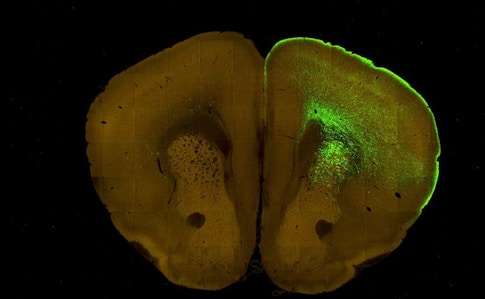
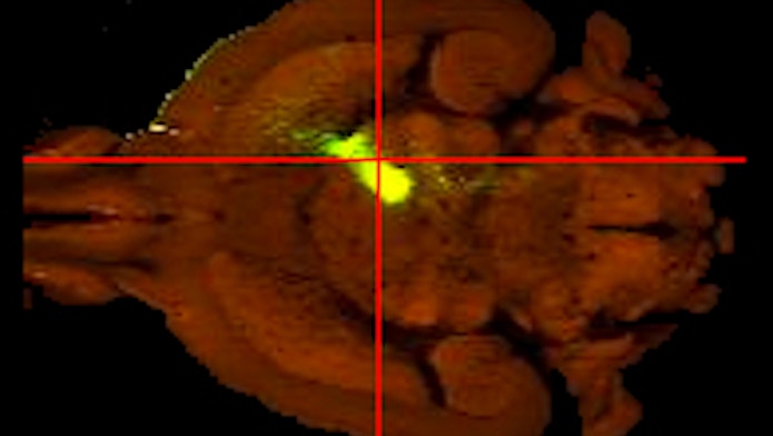
Svoboda and collaborators hadn’t set out to study the thalamus. They used the Allen Brain Atlas and other resources to figure out which brain regions connect to the anterior lateral medial cortex. They found four regions with reciprocal connections, including the thalamus.
They then used optogenetics, a method for precisely stimulating and silencing neural activity in subsets of cells, to analyze how activity in these regions influenced cortical activity. To their surprise, silencing the brain regions most strongly connected to the ALM cortex had little effect on cortical activity. But silencing the thalamus had a profound effect. “The thalamus stood out as being able to drive short-term memory.” The findings were published in Nature in May.
“The thalamus seems to be orchestrating what’s going on in the cortex,” says Bernardo Sabatini, a neuroscientist at Harvard and SCGB investigator who was not involved in the study. “It’s acting like a reciprocal part of the cortex — they are so strongly connected you almost have to think about them together.”
The research highlights the importance of analyzing multiple parts of the brain. “This is something systems neuroscientists haven’t tackled at all — we have focused on our favorite brain region or favorite cell type,” Svoboda says. “But when we want to understand neural activity and behavior, we need to take the entire brain into account.”
The researchers are now expanding their search. Collaborator Nuo Li, who recently launched his own lab at Baylor College of Medicine, will study how cerebellar systems are involved in maintaining short-term memory.
Svoboda’s team is also narrowing down the type of circuit mechanisms that could generate the sustained activity that underlies short-term memory. “There are about four different circuit models for how the persistent graded activity might be maintained,” Svoboda says. “We designed experiments to test one after the other, and we think we are converging on one particular mechanism.”
Two related studies examined working memory in a different part of the thalamus, the mediodorsal thalamus. The two groups analyzed working memory over very different time scales — from half a second to 60 seconds — but both found that this part of the thalamus was essential for maintaining working memory.

One group described its study in Nature in May. Michael Halassa, a neuroscientist at New York University, and collaborators trained mice to respond to a visual or auditory cue after a delay. The researchers showed that the brain encodes each rule — auditory and visual — separately through a population of neurons in the prefrontal cortex. However, the neurons needed input from the mediodorsal thalamus to maintain the memory over time.
In a similar study published in Nature Neuroscience in May, Christoph Kellendonk, a neuroscientist at Columbia University, Joshua Gordon, director of the National Institute of Mental Health, and collaborators found that the mediodorsal thalamus is not necessary for encoding a spatial memory task. But it is essential for maintaining the memory. “For the maintenance of working memory, the cortex needs mediodorsal inputs,” Kellendonk says.
“These papers are among the first to elucidate an interesting role for structures like the mediodorsal nucleus — without it, the cortex is quite dysfunctional,” says Sabine Kastner, a neuroscientist at Princeton University.
Taken together, the three papers highlight a new role for thalamus in memory that spans different systems. “This is a clear demonstration that in three different tasks with three different forms of working memory, the thalamus provides a signal to the cortex that’s essential for maintaining that memory,” Sabatini says. “That in itself is a big deal.”
However, the thalamus’s precise role in working memory differed from study to study, perhaps because the studies focused on different parts of the thalamus and cortex. (They also used different optogenetics techniques to control neural activity.) Svoboda’s study suggests the anterior lateral motor cortex and parts of the thalamus form a reciprocal loop — when activity in one area is inhibited, activity in the other area collapses. The two mediodorsal thalamus studies have different results, however. In the Halassa study, inhibiting the thalamus lowered activity in the prefrontal cortex but only during the task. “Our interpretation is that the thalamus changes the mode of prefrontal cortex function, making it dependent on local recurrence,” Halassa says. In the Kellendonk study, inactivating the thalamus had little effect on overall cortical activity, but it did change the information encoded in that cortical activity. “The neurons still fire more or less the same but no longer discriminate which rule to follow,” Wang says. “The discrepancy remains to be elucidated in future research.”
Both Halassa and Kelledonk theorize that the activity of the mediodorsal neurons may be functioning sort of like attention, alerting the brain where or when to be engaged. In this case, thalamic neurons may not carry specific information about the rules of the task or what’s stored in short-term memory, but rather signal the need to maintain cortical activity and stay engaged in the task.
Beyond the implications for basic research, the findings might have clinical utility. Both Kellendonk and Halassa found that stimulating the thalamus during the task improves working memory. Previous research has found deficits in thalamic-cortical connections in schizophrenia and other disorders. And damage to the thalamus can render some people essentially unconscious, Svoboda says. In rare cases, stimulating the thalamus or surrounding areas can rouse the injured person back to consciousness. “You could imagine this as a strategy of improving memory for people with deficits in working memory, like in schizophrenia,” Kellendonk says.

