New Model Shows How Cilia Make Waves
Nearly 350 years ago, Dutch scientist Antonie van Leeuwenhoek peered into a vial of lake water through a self-made magnifying lens. Through this rudimentary microscope, he saw what he described as “animalcules with incredibly thin little feet that moved very nimbly.” The animalcules were a single-celled organism; the ‘feet’ were what we now call cilia: thousands of tiny, hairlike projections that oscillate on the surface of many cells, creating waves that can both propel the cell forward and move fluid past a stationary cell. Such motile cilia play important roles in the development and health of organisms: They create waves that pump fluid in the brain, for example, and help clear particles trapped in airways. Despite their importance, much remains unknown about the physics of how they function, especially collectively. A recently published study in the Proceedings of the National Academy of Sciences provides a new look at how ciliary waves come to be, by developing a model that incorporates the complex biology at work from the nanometer-sized motors that power cilia to the millimeter-scale waves the cilia create.
“The exciting part here was bridging the gap between microscopic detail and behavior at a larger scale,” says Brato Chakrabarti, a research fellow at the Flatiron Institute’s Center for Computational Biology (CCB) and first author on the study.
“This is the first model that looks at how the microscopic motion of motors powers the cilia to produce waves that are orders of magnitude larger,” adds co-author Michael Shelley, CCB director and head of the biophysical modeling group.
The interior of a cilium resembles a microscopic engine. Specialized nanometer-sized proteins called dyneins slide internal pieces of the cilium past each other, creating bending and movement with a periodic beat. This beating movement synchronizes across the hundreds to thousands of cilia spread over a surface to generate waves that travel through the cilia — like an invisible finger running along blades of grass. These waves can travel for distances of up to several millimeters, a huge distance compared to the cilium’s microscopic size — the equivalent of a standard 10-foot oar creating a wave that travels the length of 12 Olympic-sized swimming pools.

By clicking to watch this video, you agree to our privacy policy.
The mighty power behind these tiny cilia has long been observed. The questions for researchers have been: How exactly do groups of cilia coordinate their motions to collectively produce the waves that are so important in biological processes? And what factors influence the formation and propagation of such waves? In this new study, scientists created a biophysical model that identified four factors crucial for the emergence of ciliary waves and their powerful pumping flows. At the smallest scale, the model first considered the spontaneous oscillation of a single cilium driven by its dynein motors. Next, it took into account the interactions between cilia and the surrounding fluid, and then the interactions between neighboring cilia, which tend to repel each other. Finally, the model examined how the arrangement of cilia on a surface impacted wave formation. “Physicists like simple conceptual models, but biology is complex, and this model incorporates that complexity,” says study co-author Sebastian Fürthauer, until recently a research scientist in Shelley’s group at the CCB, and now at the Institute of Applied Physics at TU Wien, the Vienna University of Technology.
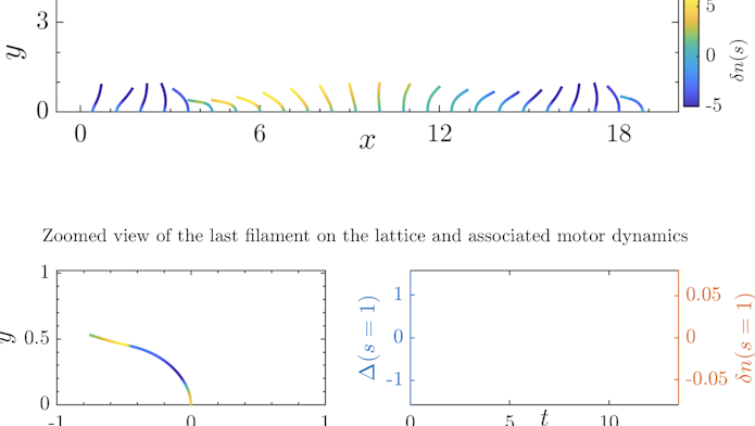
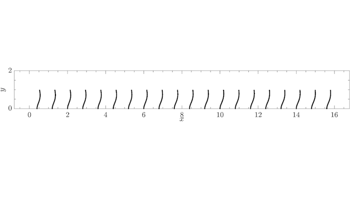
The resulting biophysical model not only showed the contributions of these four factors to the formation and propagation of ciliary waves, but also showed — in a surprise to the researchers — situations in which waves wouldn’t appear. Indeed, one unexpected finding was that sometimes a perfect, homogeneous one-dimensional row of cilia, known as an idealized lattice, produced waves, and at other times it did not. “In many cases, cilia in these idealized lattices beat in phase, and we did not see waves,” says Chakrabarti.

By clicking to watch this video, you agree to our privacy policy.
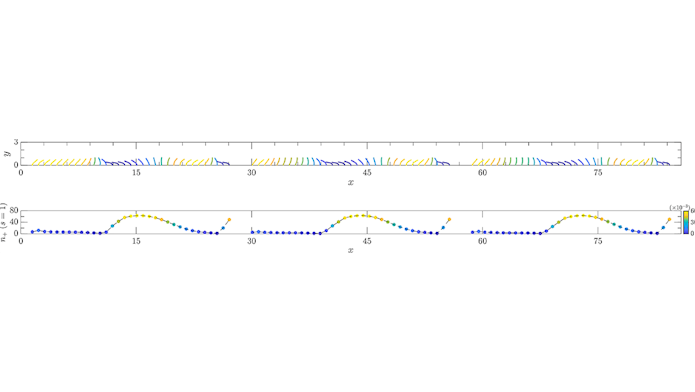
However, unlike in the scientists’ model, in nature idealized lattices don’t exist; groups of cilia are always imperfect. For example, cilia often appear in patches across the cellular surface, rather than in a uniform carpet. Sometimes this patchiness confers an advantage, such as more effective particle clearing in the airway of a mouse, as studies have shown. To explore this idea further, the researchers incorporated patchy ciliary beds into the model and saw waves emerge robustly in each of the patches and coordinate over time across the ciliated patches. “Biology says, ‘Give me a mess, and I’ll give you waves,’” says Shelley.
“Patches prevent the system from getting stuck in one kind of wave, and give flexibility. We see that what happens at the patch boundaries shapes how the waves look,” says Fürthauer.

By clicking to watch this video, you agree to our privacy policy.
The modeling here was primarily done on a one-dimensional system, but early results show the model’s consistency in two dimensions as well. Future work will focus on developing the two-dimensional modeling, taking into account real-world characteristics like uneven cilia spacing and deviations in beat frequency. Ultimately, says Fürthauer, simulating a whole organ, like a brain or a lung, will be important in understanding the impact of ciliary wave dynamics on a biological system.
Just as Leeuwenhoek appreciated the order of cilia as he gazed through his lens into a sea of complexity, today’s biophysical modelers seek to find the order in a biological system without dampening its intricacies. This is the art of biophysical modeling — uncovering the order within the complexity without getting caught in what Shelley calls the “mathematical model mindset.” “In the model, we learned that we can’t make the cilia so uniform but [can] make them more like what we see in nature, and then we can understand what nature uses to get waves,” he says.
“This work shows what you can do with a complex biophysical model,” says Fürthauer.


