Visualizing Apoptosis
These pictures, from Dr. Roland Eils of the University of Heidelberg and the German Cancer Research Center, illustrate the process of programmed cell death, called “apoptosis,” using fluorescence microscopy. Dr. Eils’ work combines mathematical modeling with experiments in molecular cell biology to yield a detailed, quantitative understanding of basic cellular mechanisms. His knowledge in the fields of physics, mathematics and biology enables scientific results not likely attainable through a traditional approach. Such an integration of expertise comprises the relatively new field of systems biology, an illustration of this report’s emphasis on cross-disciplinary activities.
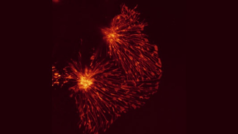
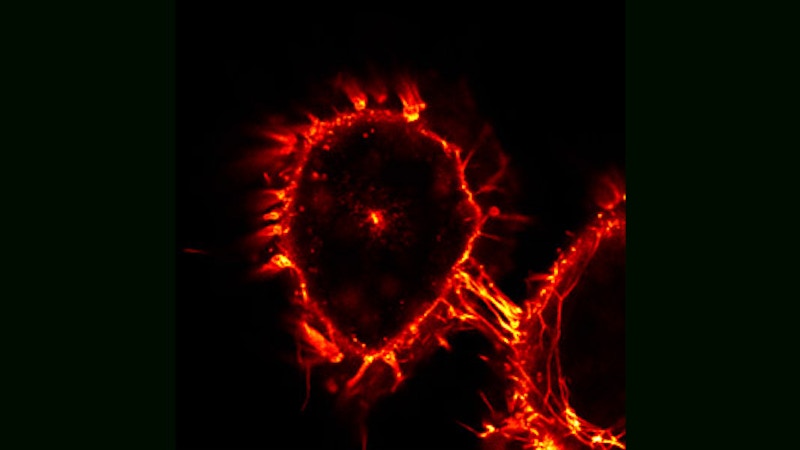
Topology of Cell Death
Here, two HeLa cells, derived from cervical cancer, are shown that have been triggered for cell death by an extracellular soluble CD95 death ligand. As soon as the CD95 death receptor is stimulated, cells start the process of apoptosis. Typically cells die within a few hours depending on the strength of cell death induction. As the cells die, they become rounder, while still keeping membrane contacts on the substrate.
Reorganization of Death Receptors
CD95 receptor activation by its CD95 ligand induces a reorganization of the receptor, including clustering and internalization. Those processes are involved in the transduction of the apoptotic signal. Here, a HeLa cell expressing CD95 fused to a fluorescent protein and induced by an extracellular soluble CD95 ligand has been imaged. The receptor is mostly present on the plasma membrane of the cell but can also be seen internalized inside the cell.
Spatial Organization of Death Receptors
Imaged here are HeLa cells; the dotted pattern shows the fluorescently tagged ligand for the receptor CD95, the molecule CD95L, which triggers apoptosis in target cells. While the CD95 receptor is present in most cells of our body, the CD95 ligand is expressed only in specialized cells and kept trapped inside these cells until they are required to kill target cells.
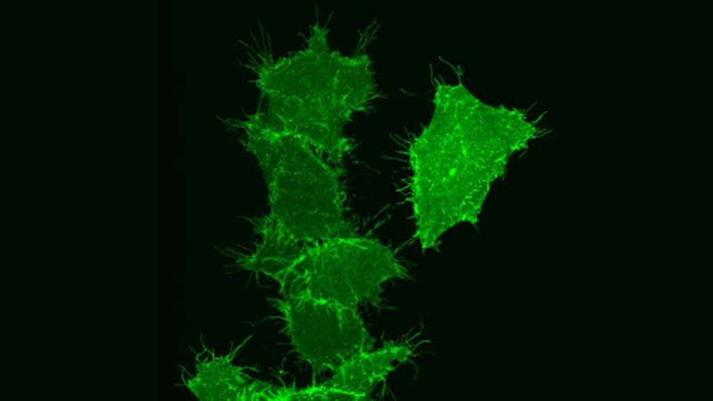
Dying Cancer Cell
The cervical cancer cell shown here is undergoing ‘apoptosis,’ programmed cell death, which occurs when the CD95 death receptor on the surface of the cell is triggered by its ligand, the molecule CD95L.
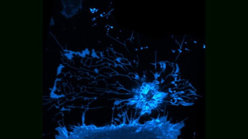
Heterogeneous Distribution of Death Receptors
CD95 receptors fused to fluorescent proteins are typically homogeneously distributed over the plasma membrane. Here, HeLa cells expressing such receptors are imaged through fluorescence microscopy by focusing on the substrate on which the cells adhere. The heterogeneities that can be seen may correspond to the complex topology of the membrane, but they may also be attributed to local accumulations of receptors in domains such as focal adhesion sites.
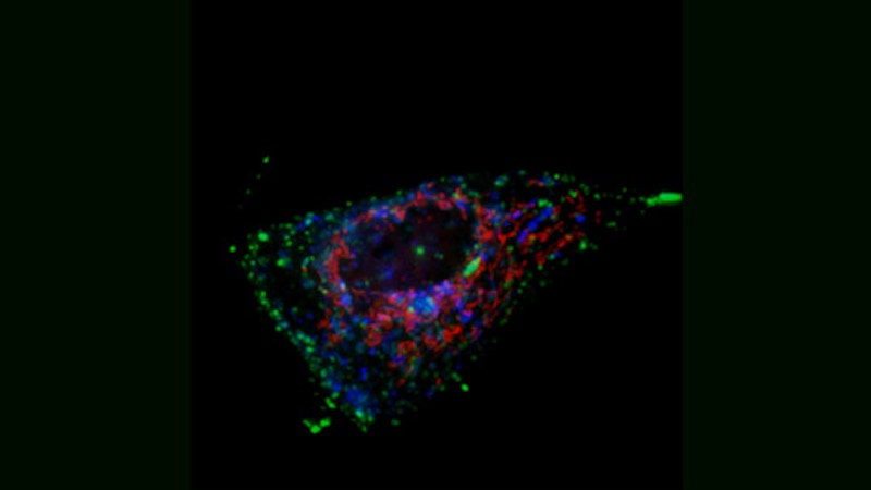
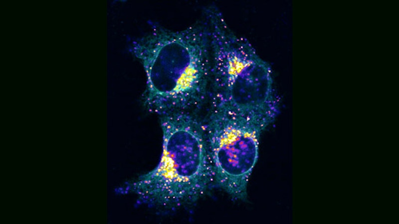
Power Plants and Transport Containers
In this breast cancer cell, fluorescence microscopy reveals three important cellular structures: mitochondria (red), early endosomes (green), and late endosomes/lysosomes (blue). Mitochondria are membrane-enclosed organelles that have a diameter of a few hundred nanometers, and lengths of up to several microns. Mitochondria are considered the cellular power plants since they produce most of the ATP that is used as a source of chemical energy, but they are also involved in a number of other essential cellular processes, such as cell death and the control of cell cycle and growth. Endosomes are membrane-bound compartments that underlie intracellular membrane trafficking. Early endosomes regulate receptor sensing of extracellular cues. Late endosomes are sites of intracellular degradation (i.e. lysosomes) and essential components of signal transduction and metabolic pathways.
Topology of Cell Death
Here, two HeLa cells, derived from cervical cancer, are shown that have been triggered for cell death by an extracellular soluble CD95 death ligand. As soon as the CD95 death receptor is stimulated, cells start the process of apoptosis. Typically cells die within a few hours depending on the strength of cell death induction. As the cells die, they become rounder, while still keeping membrane contacts on the substrate.